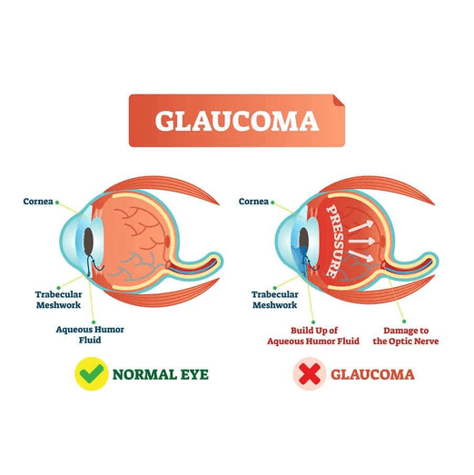
Glaucoma
Glaucoma is a group of eye conditions that damage the optic nerve, often due to increased intraocular pressure (IOP). It is one of the leading causes of irreversible blindness worldwide. Early detection and treatment are crucial to prevent vision loss. Here, we explore several diagnostic tools and treatment options used in managing glaucoma.
Automated Perimetry
Automated perimetry is a diagnostic test that measures the visual field, which is crucial for detecting and monitoring glaucoma. It assesses the patient's peripheral vision and identifies areas of vision loss.
Benefits:
- Provides a detailed map of the visual field
- Detects early signs of glaucoma-related vision loss
- Monitors disease progression and treatment effectiveness
Procedure:
- The patient sits in front of a dome-shaped device and focuses on a central point.
- Lights of varying intensities are flashed in different areas, and the patient presses a button when they see the light.
- The test results create a visual field map, highlighting any defects.
Applications:
- Diagnosis of glaucoma and other optic nerve disorders
- Monitoring progression and response to treatment
YAG Laser Treatment
YAG laser treatment, specifically YAG laser peripheral iridotomy, is used to treat angle-closure glaucoma by creating a small opening in the iris to improve fluid drainage.
Benefits:
- Quick, outpatient procedure
- Immediate reduction in intraocular pressure
- Prevents acute angle-closure glaucoma attacks
Procedure:
- The eye is numbed with anesthetic drops.
- A YAG laser is used to create a small hole in the peripheral iris.
- This allows the aqueous humor to flow more freely, reducing pressure.
Considerations:
- Used primarily for angle-closure glaucoma
- May require adjunctive treatments for long-term pressure control
Anterior Segment Optical Coherence Tomography (OCT)
Anterior segment OCT is a non-invasive imaging technique that provides high-resolution cross-sectional images of the anterior eye structures, including the cornea, iris, and angle.
Benefits:
- Detailed visualization of the anterior chamber angle
- Assists in diagnosing angle-closure glaucoma
- Guides treatment decisions for various anterior segment disorders
Procedure:
- The patient places their chin on a chin rest.
- The OCT device captures images of the anterior eye without direct contact.
- The images are analyzed to assess angle configuration and other structures.
Applications:
- Evaluation of the angle in glaucoma patients
- Assessment of corneal and lens pathologies
Retinal Nerve Fiber Layer (RNFL) Imaging
RNFL imaging is a critical diagnostic tool in glaucoma management, as it measures the thickness of the retinal nerve fiber layer around the optic nerve. Thinning of the RNFL is a hallmark of glaucoma.
Benefits:
- Early detection of glaucoma before visual field loss occurs
- Quantitative measurement of nerve fiber layer thickness
- Monitoring disease progression and response to treatment
Procedure:
- The patient looks into an OCT or scanning laser polarimetry device.
- High-resolution images are captured to assess the RNFL thickness.
- The data is compared to normative databases to identify abnormalities.
Applications:
- Diagnosis and monitoring of glaucoma
- Differentiating between glaucomatous and non-glaucomatous optic nerve damage
