Refractive Surgery
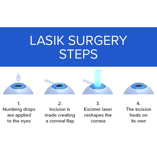
LASIK (Laser-Assisted In Situ Keratomileusis)
LASIK is the most common type of refractive surgery. It uses an excimer laser to reshape the cornea, allowing light to focus properly on the retina.
Benefits:
- Quick recovery time
- Minimal discomfort
- High success rate for correcting myopia, hyperopia, and astigmatism
Procedure:
- A thin flap is created on the cornea using a microkeratome or femtosecond laser.
- The cornea is reshaped with an excimer laser.
- The flap is repositioned and heals naturally.
PRK (Photorefractive Keratectomy)
PRK is similar to LASIK but involves removing the outer layer of the cornea (epithelium) before reshaping the underlying corneal tissue with an excimer laser.
Benefits:
- Suitable for patients with thin corneas
- No flap-related complications
- Effective for myopia, hyperopia, and astigmatism
Procedure:
- The epithelium is removed.
- The cornea is reshaped with an excimer laser.
- A bandage contact lens is placed on the eye to aid healing.
Refractive Lens Exchange (RLE)
RLE is similar to cataract surgery but performed to correct refractive errors. The natural lens is removed and replaced with an artificial lens.
Benefits:
- Effective for presbyopia and high refractive errors
- Prevents cataract development
- Offers options for multifocal or accommodating lenses
Procedure:
- The natural lens is removed using phacoemulsification.
- An artificial intraocular lens is implanted.
Corneal Topography
Corneal topography is a non-invasive imaging technique used to map the surface curvature of the cornea. It provides a detailed, three-dimensional representation of the cornea, which is essential for diagnosing and managing various eye conditions.
Benefits:
- Accurate assessment of corneal shape and refractive power
- Essential for diagnosing conditions like keratoconus and corneal astigmatism
- Aids in planning refractive surgeries and fitting contact lenses
Procedure:
- The patient looks into a device that projects concentric rings of light onto the cornea.
- The reflections are captured by a camera and analyzed to create a topographical map.
- The map provides detailed information about the corneal shape and irregularities.
Applications:
- Preoperative evaluation for LASIK and other refractive surgeries
- Monitoring of corneal diseases like keratoconus
- Assessment for contact lens fitting
Implantable Collamer Lenses (ICL)
Implantable Collamer Lenses (ICL) are a type of phakic intraocular lens implanted in the eye to correct refractive errors, such as myopia, hyperopia, and astigmatism. ICLs are an alternative for patients who are not suitable candidates for laser vision correction.
Benefits:
- Suitable for high degrees of refractive errors
- Preserves the natural lens and does not require corneal reshaping
- High-quality vision with reduced risk of dry eye
Procedure:
- A small incision is made in the cornea.
- The ICL is inserted and positioned between the iris and the natural lens.
- The incision is self-sealing and does not require sutures.
Considerations:
- Reversible procedure
- Suitable for patients with thin corneas or dry eyes
- Requires a healthy anterior chamber depth
Phototherapeutic Keratectomy (PTK)
Phototherapeutic Keratectomy (PTK) is a laser procedure used to treat corneal diseases and surface irregularities. Unlike refractive surgeries like LASIK, PTK is focused on improving corneal health rather than correcting vision.
Benefits:
- Effective for treating corneal scars, opacities, and dystrophies
- Can smooth corneal surface irregularities
- Promotes epithelial healing
Procedure:
- The epithelium is removed or modified to access the underlying corneal tissue.
- An excimer laser is used to ablate the diseased or irregular corneal tissue.
- The epithelium regenerates over the treated area.
Applications:
- Treatment of recurrent corneal erosions
- Removal of corneal scars and opacities
- Management of corneal dystrophies
